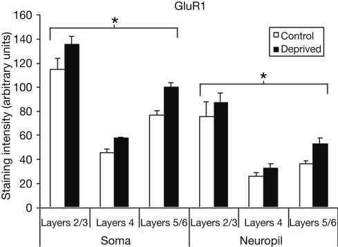
Figure 3.
Comparison of average staining intensities for GluR1 in deprived and control hemispheres. Conventions are the same as in Figure 2. GluR1 staining intensities are higher in the deprived hemisphere in all layers for both cell body and neuropil measurements (*p < 0.001).

