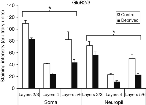
Figure 4.
Comparison of average staining intensities for GluR2/3 in deprived and control hemispheres. Conventions are the same as in Figure 2. GluR2/3 staining intensities are lower in the deprived hemisphere in all layers for both cell body and neuropil measurements (*p < 0.001).

