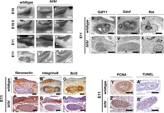
Figure 2.
In vivo bl/bl phenotypes, gene expression and cell turnover. (A)–(C), (E)–(G) and (I)–(K) are wholemounts immunostained for Pax2 marking MD, UB and both uninduced and induced MMs in black; (D), (H), (L)–(R), (Y), (Z), (A′) and (B′) were counterstained with haematoxylin. Sections in (M)–(R) and (Y), (Z), (A′) and (B′) underwent IHC (positive brown signal); sections in (S)–(X) underwent ISH (positive black signal). Wild-type E10 (A), E10.5 (B) and E11 [(C) and (D)] renal tracts showing UB initiation from the MD and growth into MM. (E)–(H) bl/bl renal tracts at E10 (E), E10.5 (F) and E11 [(G) and (H)] in which UBs fail to initiate. (I)–(L) bl/bl mutants at E10 (I), E10.5 (J) and E11 [(K) and (L)], where UBs sprout but fail to enter MM [indicated by ‘*’ in (K)]. (M)–(R) Wild-type MMs downregulated fibronectin and upregulated Itga8 and Bcl2. Mutant MMs expressed fibronectin but not significant levels of Itga8 and Bcl2. (S)–(X) Gdnf expression and Gdf11 expression were deficient in bl/bl MMs, whereas mutant MDs and UB stumps expressed Ret [arrows in (X)]. Dotted lines in (W) and (X) outline the MM. (Y) and (Z) IHC for PCNA. Note that this surrogate marker of proliferation was detected in most nuclei of wild-type and mutant MMs (brown nuclei). (A′) and (B′) TUNEL probe. Note numerous apoptotic nuclei (red) in bl/bl MM but not in wild-type MM. Bars in all frames are 100 µm.

