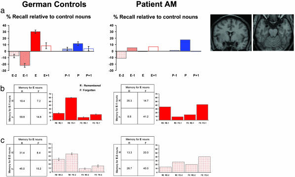
Fig. 3.
Emotion-induced memory impairment is amygdala-dependent. (a) Recall performance relative to control nouns (%) is plotted for the German control group and patient A.M. in Exp. 3. (Right) Coronal and transverse T1 MRI images demonstrating patient A.M.'s bilateral amygdala lesion are shown. A group (patient, control) × oddball type (emotional, perceptual) × position (oddball-1, oddball, oddball + 1) 2 × 2 × 3 ANOVA (assumes variance in patient population is equal to that in normals) revealed a significant group × oddball interaction (F1.0, 1.0 = 196.6 P < 0.05) and a trend for the group × oddball × position interaction (F1.0, 1.0 = 60.0 P = 0.08). A group × position 2 × 3 ANOVA restricted to the emotional factor revealed a significant group × position interaction (F1.0, 1.0 = 256.4 P < 0.05). Post hoc independent t tests revealed a significant difference between patient and controls only for recall of E-1 (P < 0.01 two-tailed) and E (P < 0.005 two-tailed) nouns. Recall of control nouns was not significantly different between patient and controls; mean recall of control nouns (%; SE) for patient A.M. 44.7; controls 47.6 (2.1). (b) Reciprocal codependency of memory for E nouns and E-1 nouns for controls and patient A.M., as for Fig. 2b. The control group demonstrates the same reciprocal effects as the placebo group in Exp. 2 (Fig. 2b). The presence of bilateral amygdala lesions abolishes this codependency, evident as a significant difference between controls (expected) and patient (observed) (χ2 = 113.8; P < 0.001). (c) The reciprocal codependency of memory for E nouns and E-2 nouns present for controls is abolished in patient A.M. (χ2 = 74.6; P < 0.001).

