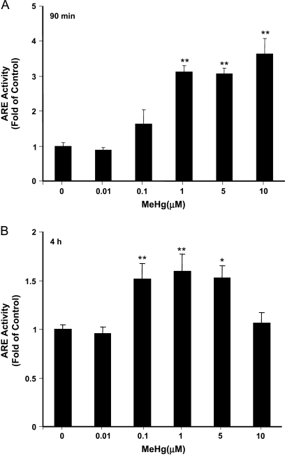
FIG. 2.
ARE activity measurement in astrocytes treated with MeHg. Cells were cotransfected with Nrf2, an ARE reporter plasmid, and a pCMV-Renilla luciferase vector for normalization. After 16 h, cells were further exposed to indicated concentrations of MeHg for 90 min (A) and 4 h (B), respectively. The ARE activities were measured by a dual luciferase assay. Each point represents the average of three separate experiments performed in duplicate (mean ± SE) (**p < 0.01; significantly different from control untreated cells; one-way ANOVA and Dunnett post hoc test).

