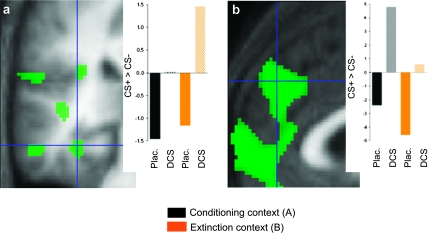
Figure 3.
Effects of postlearning DCS on fMRI correlates of recall of fear memory on day 2. Activations associated with recall of fear memory on day 2 in left posterior hippocampus/collateral sulcus (x = −34, y = −32, z = −16) (a) and right MPFC/ACC (x = 2, y = 46, z = 34) (b) were larger in the DCS than in the placebo group. Images show the parametric contrast (CS+ > CS−)DCS > (CS+ > CS−)placebo, display threshold P ≤ 0.01. Hair cross denotes activation peak surviving SVC at P ≤ 0.05. Activations are superimposed on the mean structural image. The bar graphs show average contrast estimates for the parametric CS+ > CS− contrasts in both groups and contexts.

