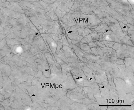
Figure 4.
Photomicrograph of DAT-ir axons (black) in the monkey VPM region lying ventral to the CM–Pf complex, and in the VPMpc. Note the presence of many thick DAT-ir axons in both nuclei that are either isolated (black arrowheads) or grouped in bundles (black arrows). Some of the thick DAT-ir axons show a ringlet-like shape (black arrowhead outlined in white). The thick DAT-ir axons, possibly representing passing axons, are mixed with the typical thin varicose axons prevalent in most other thalamic nuclei.

