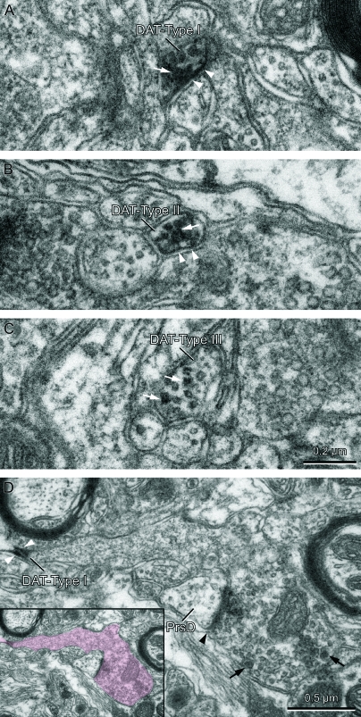
Figure 5.
Ultrastructure of DAT-ir axons in the macaque MD. (A–C) Transversely cut inmunolabeled axons showing the 3 types of labeling we identified. (D) Longitudinally cut immunolabeled axon with its terminal endowed with vesicles and a synaptic profile (identified in color in the inset). White arrows in (A)–(C) point to immunoprecipitate in the cytoplasm and axonal organelles. White arrowheads in (A), (B), and (D) point to immunoprecipitate rimming the inner border of the plasma membrane, a feature present in DAT-Type I and DAT-Type II immunolabeling patterns. DAT-Type I immunolabeling is characterized by intensely electron dense immunoprecipitate conspicuously attached to the plasma membrane (A and D), whereas DAT-Type II immunolabeling is characterized by the immunoprecipitate preferentially located within the cytoplasm and its organelles (B). In the DAT-Type III immunolabeling pattern, the immunoprecipitate is confined to the cytoplasm and mostly associated to microtubules (C). The axon in D establishes a synaptic contact (black arrowhead) that is located far from the DAT immunoperoxidase product (white arrowheads). The axon terminal contains many synaptic vesicles, some of them with dense cores (black arrows). Note the presence of vesicles in the postsynaptic element, thus identified as a presynaptic dendrite (PrsD), typical of thalamic interneurons. The calibration bar in (C) applies to (A)–(C).

