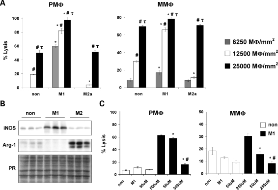
Figure 3.
Classically activated macrophages mediate muscle cell lysis via an NO-dependent mechanism. Cytotoxicity assays showed that M1 peritoneal (left panel, PMΦ) and mdx muscle macrophages (right panel, MMΦ) had the highest cytotoxic activity (A). *, P < 0.05 when compared with non-stimulated control at the same concentration. #, P < 0.05 when compared with macrophages cultured at 6250 MΦ/mm2 within the same treatment condition. τ, P < 0.05 when compared with macrophages cultured at 6250 and 12 500 MΦ/mm2 within the same treatment condition. The increase in muscle cells lysis mediated by M1 muscle macrophages paralleled increases in iNOS expression when they were cocultured with myotubes (B). Cytotoxicity assays performed in the presence of L-NAME showed that inhibition of NO synthesis resulted in a dose-dependent decrease in muscle cell lysis mediated by M1 peritoneal and muscle macrophages (C). *, P < 0.05 when compared with M1. #, P<0.05 when compared with M1 treated with 50 µM L-NAME. Non, non-stimulated macrophages; M1, M1 macrophages; M2a, M2a macrophages; PR, nitrocellulose membranes stained with ponceau red to ensure equal loading of total protein. Representative histograms or blots of 2–3 independent experiments are shown.

