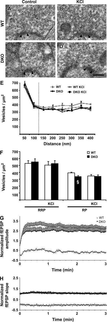
Figure 6.
Effects of F-actin modulation on vesicle densities and frequency facilitation at MF–CA3 synapses in slices. (A) Electron micrograph from a control WT MF terminal (t) exposed to cytochalasin B (20 μM). The synapse is indicated by its postsynaptic density (arrowheads), spine (s), vesicles (v), and mitochondria (m). Scale bar: 0.1 μm. (B) As in (A), but the electron micrograph is from a WT MF terminal which subsequently to cytochalasin B (20 μM) exposure was stimulated with 30 mM [KCl]o. (C, D) As in (A) and (B), but the electron micrographs are from synapsin DKO MF terminals. Artifactual small, electron-dense precipitates are present in electron micrographs (A–D). (E) Bar graph (mean + standard error of the mean [SEM]) showing the vesicle densities (vesicles/μm2) at increasing distance from the presynaptic membrane specialization in both genotypes in control situation and exposed to high [KCl]o. (F) Bar graph results are grouped into distances of 0- to 100-nm distance and 0–400 nm, corresponding to RRP and RP, respectively. Vesicle densities were compared between genotypes at the given distances and no differences found. For each of the treatments, 3 WT and 3 synapsin DKO mice (the number of synapses in each animal ranged from 18 to 34 in 20 electron micrographs from each slice). Note the increase in DKO vesicle density, rather than a decrease in WT. §P < 0.05 between DKO with and without cytochalasin B when comparing Figure 2F. (G) Normalized and pooled MF-elicited fEPSPs in WT (open circles) and synapsin DKO (filled circles) mice following the switch from 0.1- to 2-Hz stimulation (only the first 3 min are shown) in the presence of cytochalasin B (20 μM). Subtraction of the values obtained in synapsin DKO mice from those obtained in WT mice is represented by the open triangles. Vertical bars indicate SEM. (H) As in (F), but measurements are from responses elicited by stimulation of the AC collateral–CA3 pathway in the presence of cytochalasin B (20 μM).

