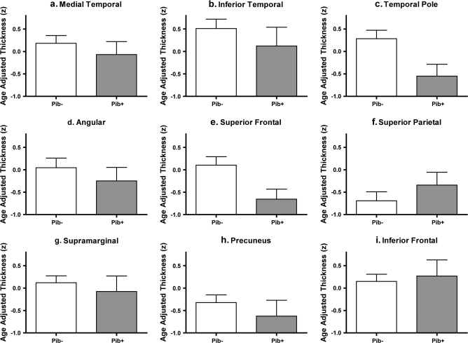
Figure 7.
Consistent subtle thinning appears to be present in many cortical ROIs in amyloid (PIB)-positive OC (CDR = 0) compared with amyloid (PIB)–negative controls. ROIs were generated from exploratory analysis in subject Sample 1a (see Fig. 2, same ROIs as used in Fig. 3) and applied to a new sample of PIB+ and PIB- controls (CDR = 0, Sample 1c) to test the hypothesis that these regions undergo subtle thinning in asymptomatic AD. Bars represent mean thickness of each region shown in Figure 2, normalized for age and standardized to Z scores. Error bars indicate 1 standard error of the mean. (A) Medial temporal cortex, (B) Inferior temporal gyrus, (C) Temporal pole, (D) Angular Gyrus, (E) Superior frontal gyrus, (F) Superior parietal lobule, (G) Supramarginal gyrus, (H) Precunes, (I) Inferior frontal sulcus.

