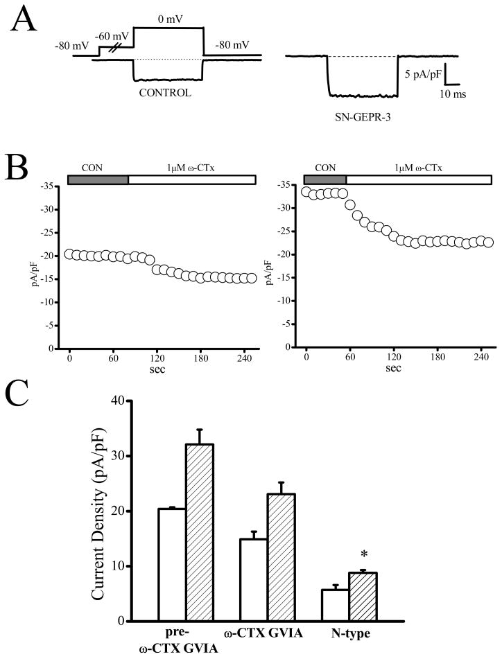
Figure 6.
Enhancement of N-type current density in IC neurons of SN-GEPR-3s. Ba2+ currents were activated at 0 mV in absence and presence of 1 μM ω-conotoxin GVIA in IC neurons obtained from control SD rats and SN-GEPR-3s. A. Representative ω-conotoxin GVIA-sensitive current trace (obtained as described in Figure 4A) in control SD rat (left trace) and SN-GEPR-3 (right trace). B. Time course of the suppressive effect of ω-conotoxin GVIA on HVA Ca2+ channel currents in control SD rat (left panel) and SN-GEPR-3 (right panel). C. The ω-conotoxin GVIA -sensitive current was larger in SN-GEPR-3s (n = 8) compared to control SD rats (n = 9). *P <0.05.

