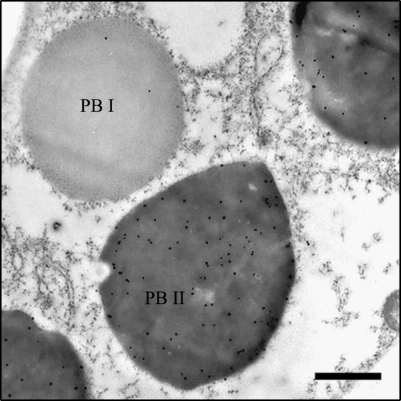
Fig. 7.
Intracellular localization of GluD-1 in the maturing seeds detected by immunoelectron microscopy. PB I and the high-density, irregularly shaped PB II are indicated. Twenty-five-nanometer (25 nm) gold particles labelled with anti-GluD-1 antibody are predominantly distributed in PB-II. Bars=1μ m.

