Fig. 1.
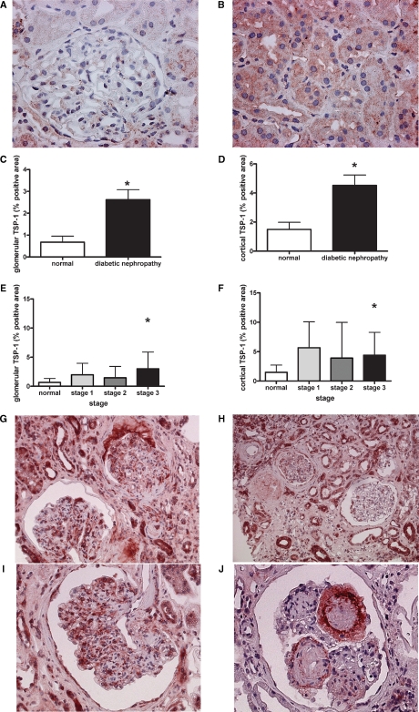
TSP-1 expression is enhanced during type-2 diabetic nephropathy. Immunohistochemistry of TSP-1 in normal kidneys demonstrated the wide absence of TSP-1 in glomeruli (A) but some low level TSP-1 in tubules (B). Glomerular (C) and cortical (D; representing mainly tubulointerstitial) TSP-1 expression were evaluated as described in the Material and methods section and were increased in type-2 diabetic nephropathy. Expression levels of glomerular (E) and cortical (F) TSP-1 varied among biopsies with different severity of lesions. With ongoing disease (G) TSP-1 expression within glomeruli could be detected in various glomerular and inflammatory cells (H, I). In the tubulointerstitium, TSP-1 was mainly confined to tubules and inflammatory interstitial cells (G, H). Kimmelstiel–Wilson lesions demonstrated a typical staining pattern (J) (*P < 0.05 by the Mann–Whitney U-test).

