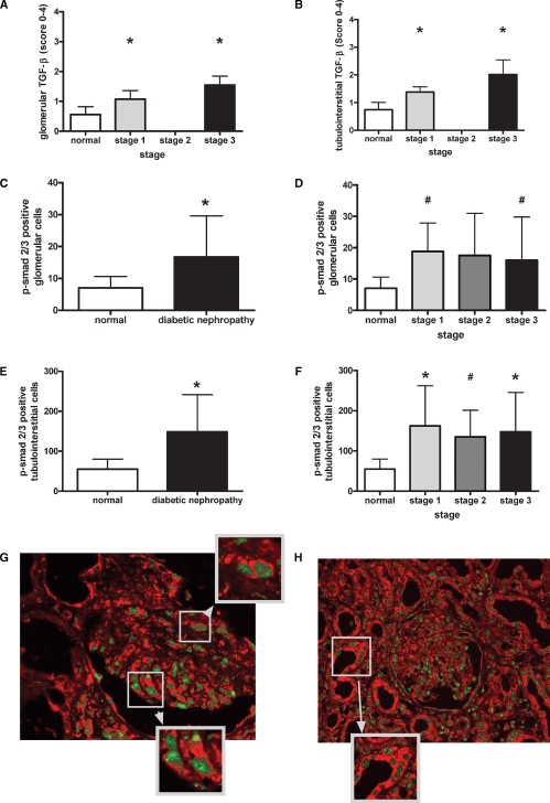
Fig. 2.
Increased TGF-β expression and p-smad2/3 positivity during advanced diabetic nephropathy. Glomerular (A) and tubulointerstitial (B) immunohistochemistry of TGF-β-1/2 were evaluated using a semiquantitative scoring system as described in the Material and methods section. Immunohistochemistry of p-smad2/3 was evaluated by counting positive cells in glomeruli and the tubulointerstitium. Glomerular p-smad2/3 was increased in all diabetic biopsies (C) and biopsies with mild (grade 1) and advanced (grade 3) lesions (D) whereas cortical p-smad2/3 was increased in all subgroups (E, F). Double labelling of TSP-1 and p-smad2/3 was performed as described in the Material and methods section (G, H). Panel G and its zoomed areas depict glomerular co-localization; panel H depicts the clear co-localized TSP-1 and p-smad 2/3 in the tubulointerstitium of diabetic kidneys (*P < 0.05 by the Mann–Whitney U-test; #P < 0.05 by the unpaired t-test and Welch-test).

