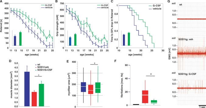
Fig. 4.
G-CSF improves motor function in SOD1-tg mice. SOD1 transgenic mice were treated with subcutaneous G-CSF (30 µg/kg/day) or vehicle (vehicle, n = 17; G-CSF, n = 18) starting at week 11 of age, where signs of muscle denervation are already obvious on EMG. The drugs were continuously administered for 8 weeks with an osmotic minipump (Alzet 2004) that was implanted in a paravertebral position at 11 weeks of age and exchanged at week 15. (A) Rotarod performance. Graph depicts means ± SEM over 14 weeks. Inset: AUC, bar graph depicts area under the curve analysis of the individual performance curves. (B) Grip strength. Graph depicts means ± SEM over 14 weeks (mN). Inset: AUC, bar graph depicts area under the curve analysis of the individual performance curves. There is considerable improvement in both motor parameters in the treated group (∼40% by AUC analysis; P < 0.05). (C) Kaplan–Meier analysis of the time passed until rotarod performance dropped below 50% of the initial value. Inset: AUC analysis. There is a significant difference between vehicle and G-CSF treatment in all parameters shown (log-rank test; P < 0.05). (D–G) G-CSF diminishes denervation atrophy in hind limb muscles of SOD1-tg mice. Shown are (D) the diameter of the M. rectus femoris of the M. quadriceps group in wt, vehicle-treated and G-CSF-treated SOD1-tg mice. There is a significant increase in the muscle diameter by G-CSF treatment. (E) Analysis of individual fibre diameters in the quadriceps group. There is a significant increase in fibre diameter by G-CSF treatment compared with the vehicle-treated group. Shown is a box-blot with median (white) and the 25% and 75% quartiles as box. (F and G) Quantification of single fiber potentials by electromyography of the M. gastrocnemius in wt, vehicle- and G-CSF-treated SOD1-tg mice at week 14. There is a significant decrease in fibrillations by G-CSF treatment. (F) Maximal frequency of fibrillation. (G) Exemplary traces from the wt, vehicle- and G-CSF-treated SOD1-tg mice at week 14 (*P < 0.05).

