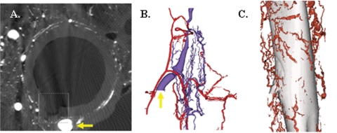
Fig. 10.
Three-dimensional microscopic computed tomographic evaluation of the carotid artery and jugular vein. (A) A pig carotid artery cross section showing the lumen and the origin of a vasa vasorum externa with microfilm (box). Yellow arrow is shown in (B). (B) The arterial vasa vasorum (red) and the venous vasa vasorum (blue) with the yellow arrow on the vasa vasorum from (A). (C) The vasa vasorum surrounding the vein that communicates with the arterial vasa vasorum.

