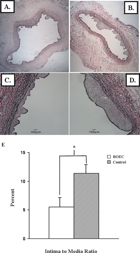
Fig. 5.
Verhoeff's van Giesen staining was performed at the venous stenosis (section V1, see Figure 1C) from the cushioning region of the BOEC-transplanted (A and C) and contralateral non-transplanted (control) veins (B and D). A and B are 5× and C and D are 40× magnification. The lower panel (E) shows that there was a 50% decrease in the intima-to-media ratio in the BOEC-transplanted samples when compared to controls (P < 0.05). Data are mean ± SD.

