Fig. 5.
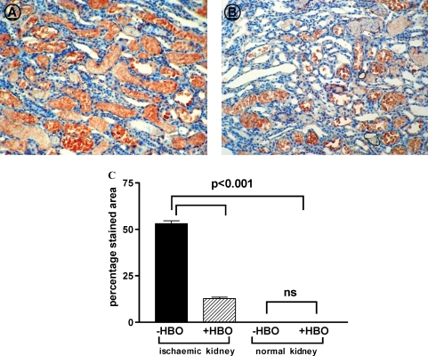
Representative pictures of 4-HNE staining in the medulla of −HBO (A) and +HBO (B) ischaemic kidneys (objective ×20). (C) Percentage staining area of 4HNE in the renal outer strip of the outer of −HBO and +HBO of control and ischaemic kidneys. n = 6–9 in each group. This figure indicates that 4-HNE is localized primarily in the renal tubular cells of the outer stripe of the medulla of the ischaemic kidneys, and is almost undetectable in the cortex. The area of the outer stripe of the medulla stained for 4-HNE is significantly less than in the ischaemic kidneys of rats treated with hyperbaric oxygen than in untreated rats.

