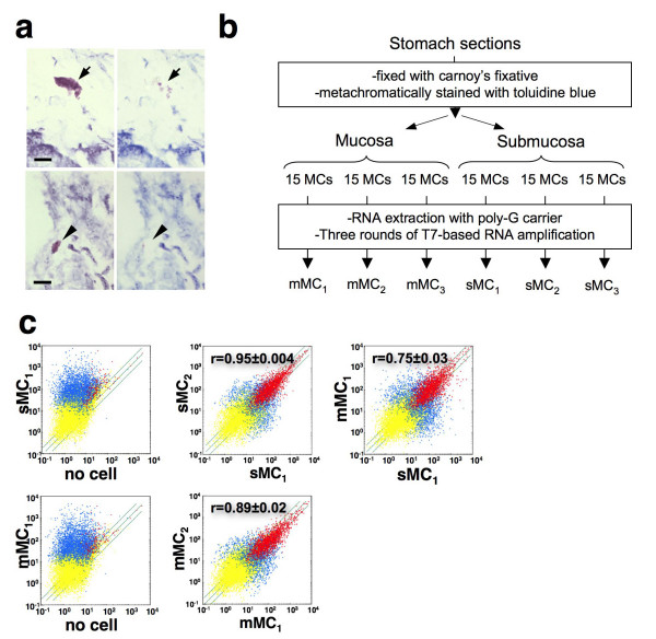
Figure 2.
Gene expression profiles of sMCs and mMCs from stomach tissue. (a) Isolation of toluidine blue-stained MCs in the submucosa (sMC; upper panels) and the mucosa (mMC; lower panels) of stomach sections. A sMC (arrow) and mMC (arrowhead) that was metachromatically stained with toluidine blue before microdissection (left panels) disappeared after microdissection with a patch pipette (right panels). Bars, 10 μm. (b) Outline of the experimental strategy. (c) Labeled and fragmented antisense RNAs of three individual sMC samples, three individual mMC samples and the 'no cell' samples were hybridized to a Murine Array. Scatter plots for 'no cell' (x axis) and sMC1 (y axis) (upper left), 'no cell' (x axis) and mMC1 (y axis) (lower left), sMC1 (x axis) and sMC2 (y axis) (upper center), mMC1 (x axis) and mMC2 (y axis) (lower center), sMC1 (x axis) and mMC1 (y axis) (upper right) are shown. The correlation coefficients (r) for comparison within sMC1–3, within mMC1–3 and between sMCs and mMCs are presented as means ± S.D. Red dots show probe sets judged as "Presence", and yellow dots represent probe sets judged as "Absence" in both arrays. Blue dots show probe sets judged as "Presence" only in either array. The same, two-fold induction and suppression thresholds are indicated as diagonal lines.

