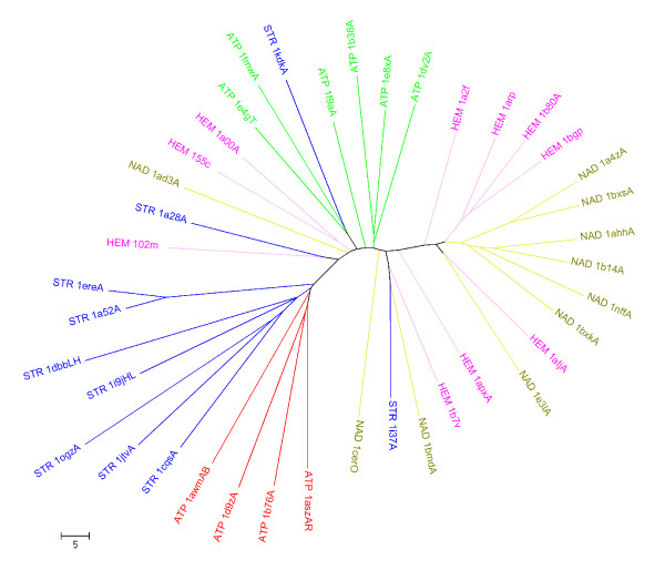
Figure 8.
A cluster tree of the 40 binding sites using the dataset of [16]. The PDB codes are prefixed by ligand codes-STR indicating a steroid, ATP, NAD and HEM referring to heme. The chain IDS where appropriate are suffixed to the PDB codes. The proteins are coloured based on their ligand types. ATP sites are shown in two colours to reflect the two different types of sites.

