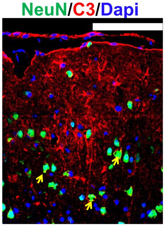
Figure 1.

Up-regulation of C3 in neurons as well as other brain cells in infarcted brain regions following ischemic stroke. Staining and counterstaining with nuclear marker DAPI indicate colocalization of the neuronal nuclear marker NeuN (green fluorescence) and membranous type of C3 immunoreactivity (yellow arrows), whereas nonneuronal brain cell exhibit cytoplasmic type of C3 up-regulation.
