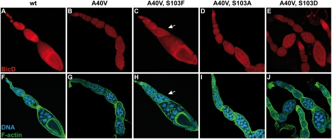
Figure 3. A S103F substitution in BicDA40V suppresses the BicDPA66 phenotype.
Confocal images of ovaries from the indicated wt and mutant females, stained with anti-BicD antibodies (red). Blue: DNA, green: F-actin. A, F: In ovaries with wild type BicD, all egg chambers contain an oocyte, and the protein accumulates in the oocyte throughout oogenesis. The BicD mutants A40V (B, G) fail to form an oocyte, and egg chambers contain 16 nurse cells. The mutant protein does not accumulate in a single cell. C, H: Substitution of Ser103 by phenylalanine in BicDA40V suppresses the BicDPA66 phenotype. Most egg chambers form an oocyte, where the BicD protein accumulates to a certain extent. The egg chamber indicated by an arrow contains 16 polyploid nurse cells and no oocyte. D, I: The mutants A40V+S103A and A40V+S103D (E, J) also fail to form an oocyte. Shown are maximum projections of z-stacks. Panels F–J: for clarity reasons, individual panels are composed of different optical sections.

