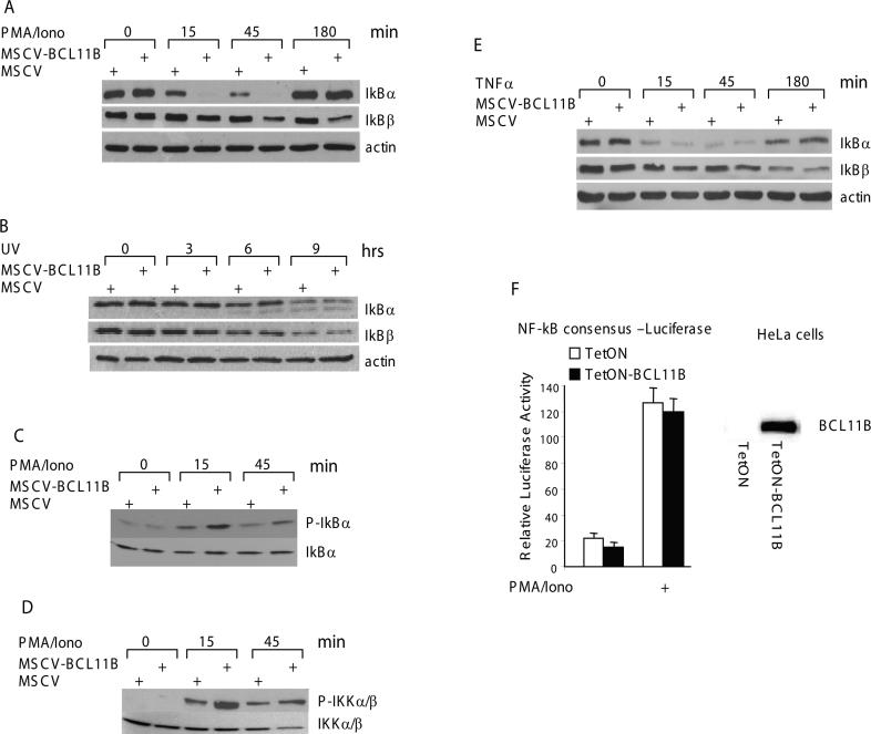
Figure 5. BCL11B enhances IkB phosphorylation and degradation, and IkB kinase phosphorylation in a TCR-dependent manner.
(A) MSCV and MSCV-BCL11B Jurkat cells were treated with 50 ng/ml PMA and 1 μM ionomycin for the indicated time points. IkB degradation was analyzed by Western blot analysis of cytoplasmic fractions with specific antibodies. Actin was used as loading control. (B) Same as in (a) except that the cells were irradiated with 80 J/m2. (C) MSCV and MSCV-BCL11B Jurkat cells were pretreated with 2.5 μM MG132 for 16 hrs followed by stimulation with 50 ng/ml PMA and 1 μM ionomycin for the indicated time points and P-IkBα was recognized with specific antibodies. The same membrane was stripped and total amount of IkBα was detected by reprobing with an anti-IkBα antibody. (D) MSCV and MSCV-BCL11B Jurkat cells were treated with 50 ng/ml PMA and 1 μM Ionomycin for the indicated time points. The P-IKKα/β were detected with specific antibodies. The total amount of IKKα/β proteins is shown on the same membrane. (E) MSCV and MSCV-BCL11B Jurkat cells were treated with 20 ng/ml TNFα for the indicated time points. IkB degradation was analyzed by Western blot analysis of cytoplasmic fractions with specific antibodies. Actin was used as loading control. (F) (Left panel) HeLa cells ectopically expressing BCL11B (solid bars) or not (open bars) were transfected with pNF-kB-Luc and Renilla Luciferase plasmids. 24 hrs posttransfection the cells were stimulated with 50 ng/ml PMA and 1 μM ionomycin for 6 hrs and the luciferase activity was evaluated. (Right panel) Western blot showing ectopic expression of BCL11B in Hela cells.

