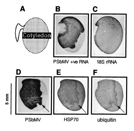
Figure 3.

Accumulation of viral and host proteins at the front of virus invasion. B and C and D–F, respectively, were cut from two different infected embryos. The orientation of the sections is illustrated in A. Sections were subjected to in situ hybridization with probes for PSbMV +ve RNA (B) or 18S rRNA (C), or immunohistochemistry with antibodies for PSbMV coat protein (D), HSP70 (E), or ubiquitin (F). rRNA was uniformly distributed throughout the infected cotyledon. Cells at the front of virus invasion (arrows) accumulated viral and induced host proteins.
