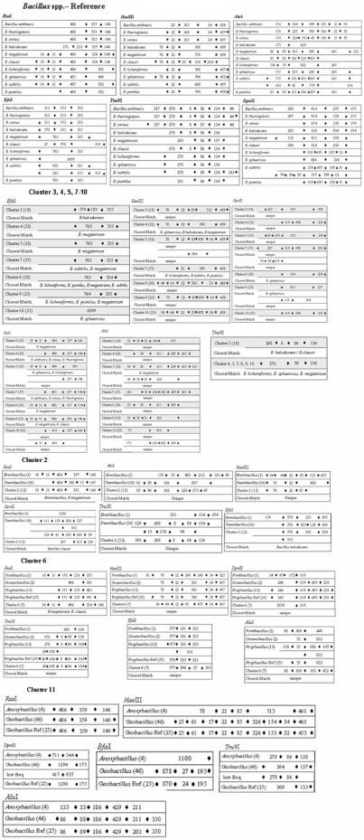
Figure 9. Nucleotide fragments generated as a result of in silico restriction enzyme (a).
RsaI (b). HaeIII (c). AluI (d). BfaI (e). Tru9I (f). DpnII, action on 16S rDNA gene sequences in 10 Bacillus spp. and 11 clusters (Figure 8). A solid black box represents the restriction enzyme cut site. At the terminal points, solid black box are shown only if highly variable fragments are observed beyond this cut site.

