
Figure 1.
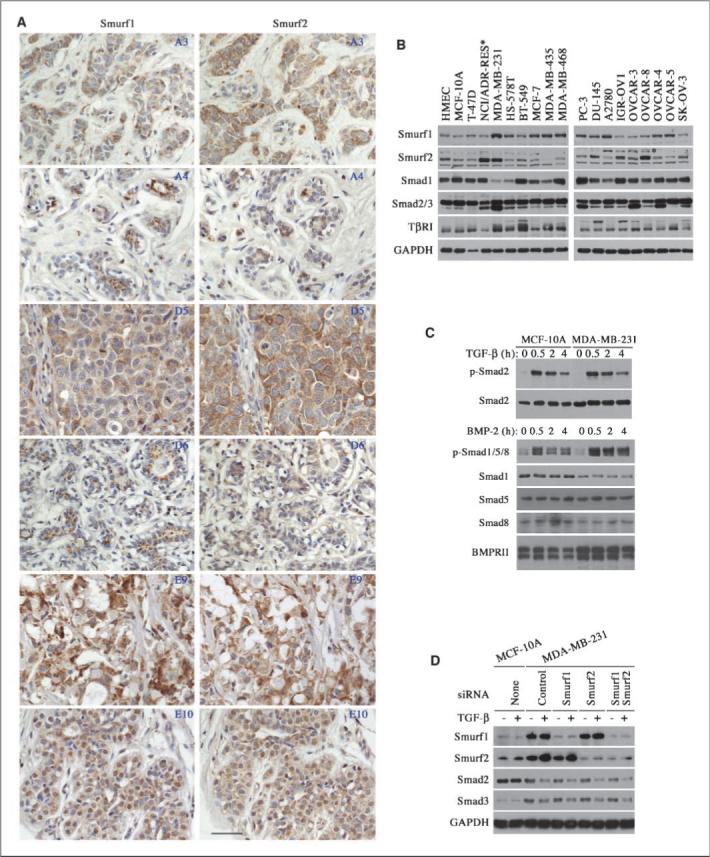
Up-regulation of Smurf2 in breast cancers. A, immunohistochemical staining of tumor (A3, D5, E9) and matching normal (A4, D6, E10) tissues in a breast cancer tissue array with anti-Smurf1 or anti-Smurf2 antibody. Three pairs of representative images are shown here and coordinates of each sample in the array are given in the top right corner. Scale bar, 4 μm. B, expression of Smurfs, Smads, and TβRI receptor in various cancer and normal control cells. *, a nonspecific band above the Smurf2 band. C, normal activation of Smads in MCF-10A and MDA-MB-231 cells. D, attenuated Smad expression induced by prolonged exposure (2 d) to TGF-β is resistant to RNAi-mediated knockdown of Smurf1 and/or Smurf2 in MDA-MB-231 cells. Left, protein levels in normal MCF-10A cells.