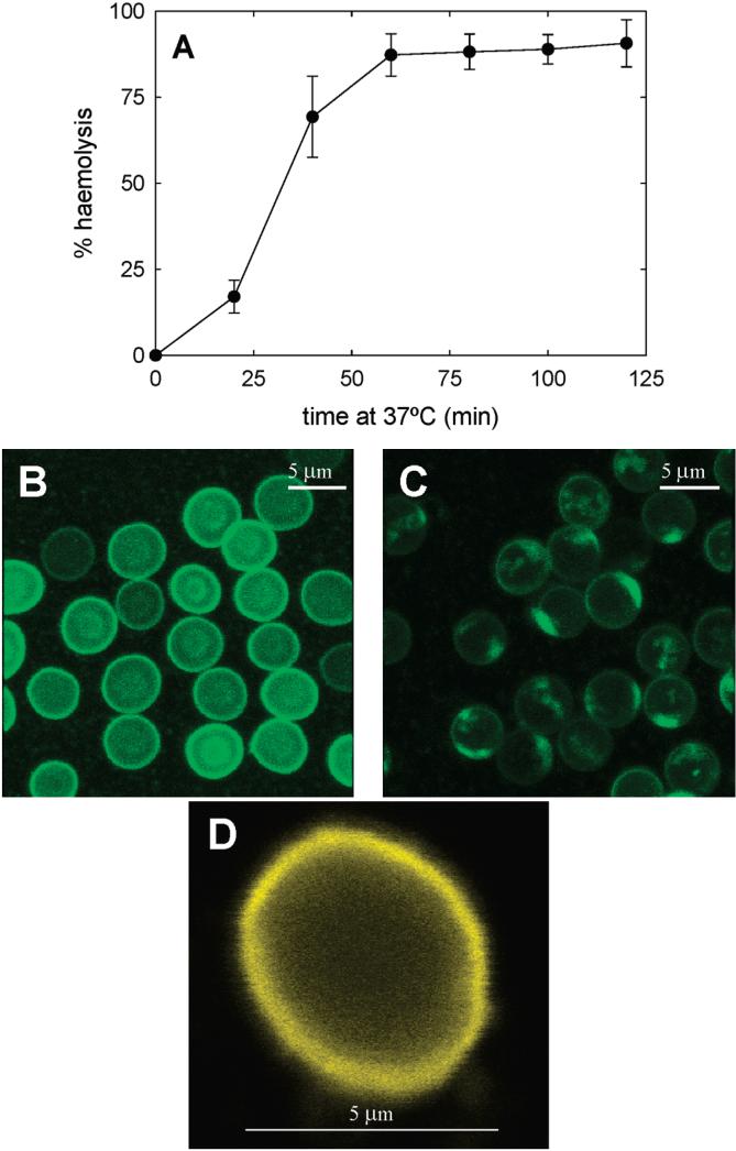
FIGURE 3.
Hot-cold hemolysis of human RBC treated with PlcHR2. (A) Time course of hot-cold hemolysis. RBC are incubated with PlcHR2 at 37 °C for varying lengths of time, as indicated on the x-axis, and then transferred to 4 °C for 2 min before quantifying the degree of hemolysis. Average values ± SD (n = 3). (B) Control RBC, examined by confocal fluorescence microscopy after staining with BODIPY FL C12-sphingomyelin. (C) RBC after 60 min incubation with enzyme at 37 °C and then cold treatment for 2 min. (D) RBC “ghosts” obtained by osmotic shock, stained with DiIC18. Bars: 5 μm.

