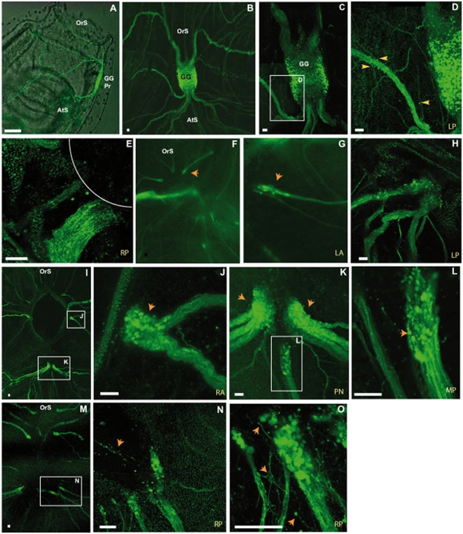
Figure 5. Cell masses arranged around nerve ends.
Nerves visualized in E15 transgenic animals. GFP is produced only after the metamorphosis of juveniles (A). Ganglion and emerging nerves in B. Nerves in close proximity to the ganglion before ablation. C and D show peripheral cell bodies (yellow arrowheads). Nerve ending just after ablation in E, with line indicating hole. At 2 dpa (stage I) no clear pattern of nerve cell bodies is observed at the nerve endings (F, G and H). Nerve ending with cell bodies forming a swelling at 4 dpa (I, arrowheads in J and arrowheads in K). Some thickened nerve cell masses start to extend their axons toward the anterior at 4 dpa (L). At 9 dpa (early stage III) the nerve end cell masses decrease in size and send lots of extensions in the anterior and posterior directions, sometimes making connections (M, arrowheads N, and O). All scale bars, 50 µm. Ganglion primordium (GG Pr), atrial siphon (AtS), oral siphon (OtS), left posterior nerve (LP), right posterior nerve (RP), mid-posterior nerve (MP), posterior nerves (PN), right anterior nerve (RA) and left anterior nerve (LA). Fluorescent confocal images obtained with the Leica SP5 system (5×, 20× and 40×).

