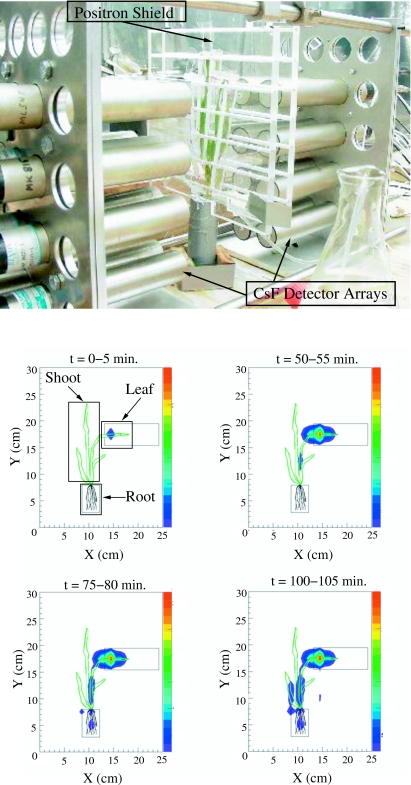
Figure 3. (Above) Photograph of a barley plant arranged within the FOV of the low spatial resolution 2D PET imager.
Each detector cylinder is a cesium fluoride scintillator with dimensions 25 mm diameter by 45 mm thick. A plastic shield equipped with ventillation ducts is placed near the plant surface to stop positrons that escape from the plant tissue so that they annihilate within the imager FOV. Individual barley leaves are also separated with plastic shields to ensure that positron annihilation occurs locally. (Below) Snapshots indicating 11C-labeled photoassimilate accumulation in a barley plant as a function of time. The integration time for these images is 5 min; the minimum exposure time is imposed by the counting rate of the detector system and the minimum required counting statistics within each region of interest. The relative intensity of the source in the image plane is coded according to the color scale on the right side of each frame with red representing the brightest pixels. The images are corrected for background radiation, radioactive decay of 11C, and the detection efficiency as a function of location in the image plane. In the first frame (0–5 min), the regions of interest (leaf, shoot, and root, as indicated by the rectangular boxes) are selected for direct comparison to earlier collimated detector measurements.

