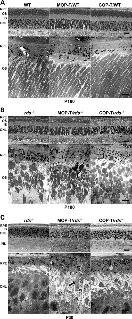
Figure 5.
C150S improves rod photoreceptor OS structure in rds+/− retina. Shown are representative light (top) and electron (bottom) microscopy from retinal sections of MOP-T, COP-T and non-transgenic controls in the WT (A), rds+/− (B) and rds−/− (C) backgrounds. Eyes presented in A and B were taken from mice at P180 and in C from mice at P30. Expression of C150S-Rds had no effect on rod OS structure in the WT background and led to partial improvement in OS structure in the rds+/− background. As expected, no change in rod OS structure was noticed in COP-T/rds+/− or COP-T/WT retinas compared with non-transgenic controls. (C) Except in rare instances (arrow) C150S-Rds was not sufficient to support OS formation in the absence of endogenous Rds. RPE, retinal pigment epithelium; OS, outer segment; IS, inner segment; ONL, outer nuclear layer; INL, inner nuclear layer. Scale bar, 4 µm (EM) and 20 µm (light).

