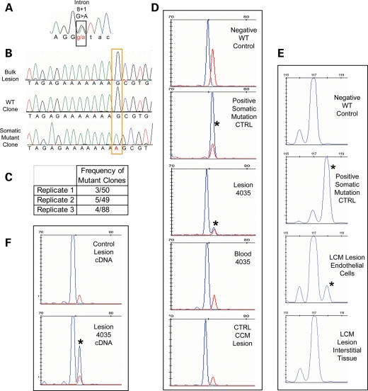
Figure 4.
A CCM3 somatic mutation identified in sample 283-4035. (A) The germline mutation identified by sequence analysis of leukocyte-derived DNA is a splicing mutation in CCM3 exon 8, c.474+1 G>A. (B) Somatic mutation identified as an insertion of A in exon 6 of CCM3, c.205-211insA. The somatic mutation is not readily identifiable from the direct sequence of bulk lesion-derived DNA. Clonal analysis reveals both wild-type and somatic mutant clones. (C) The somatic mutation is identified multiple times in each of three replicates. The final replicate is of Phusion-derived PCR products. (D) The somatic mutation is validated by fragment analysis. Size standard marker at 75 bp is in red. Phusion-derived PCR fragments appear in blue. The wild-type fragment product is 74 bp (Negative Ctrl), and the somatic mutation product is 75 bp long (Positive Ctrl). The lesion-derived sample yields two products, both wild-type and mutant (asterisk) with the mutant allele representing 10% of the total amplicons. The larger fragment of the somatic mutation is not present in either matched blood for lesion sample 4035 or another unrelated CCM lesion sample that is WT at the site of the somatic mutation. (E) In a different fragment analysis assay, normal WT length fragments are 117 bp, and those with the somatic mutation (asterisk) are 118 bp long. From the laser-captured tissue, only the endothelial cells and not the interstitial tissue harbor the somatic mutation. Thirteen percent of the endothelial cell-derived amplicons carry the somatic mutation. (F) The somatic mutation is biallelic to the germline mutation. Fragment analysis of a phusion-derived PCR product from lesion cDNA spanning exons 6 to 9 of CCM3. The somatic mutation (asterisk) is only present in the RT–PCR product from 4035 lesion cDNA, not from a control CCM lesion.

