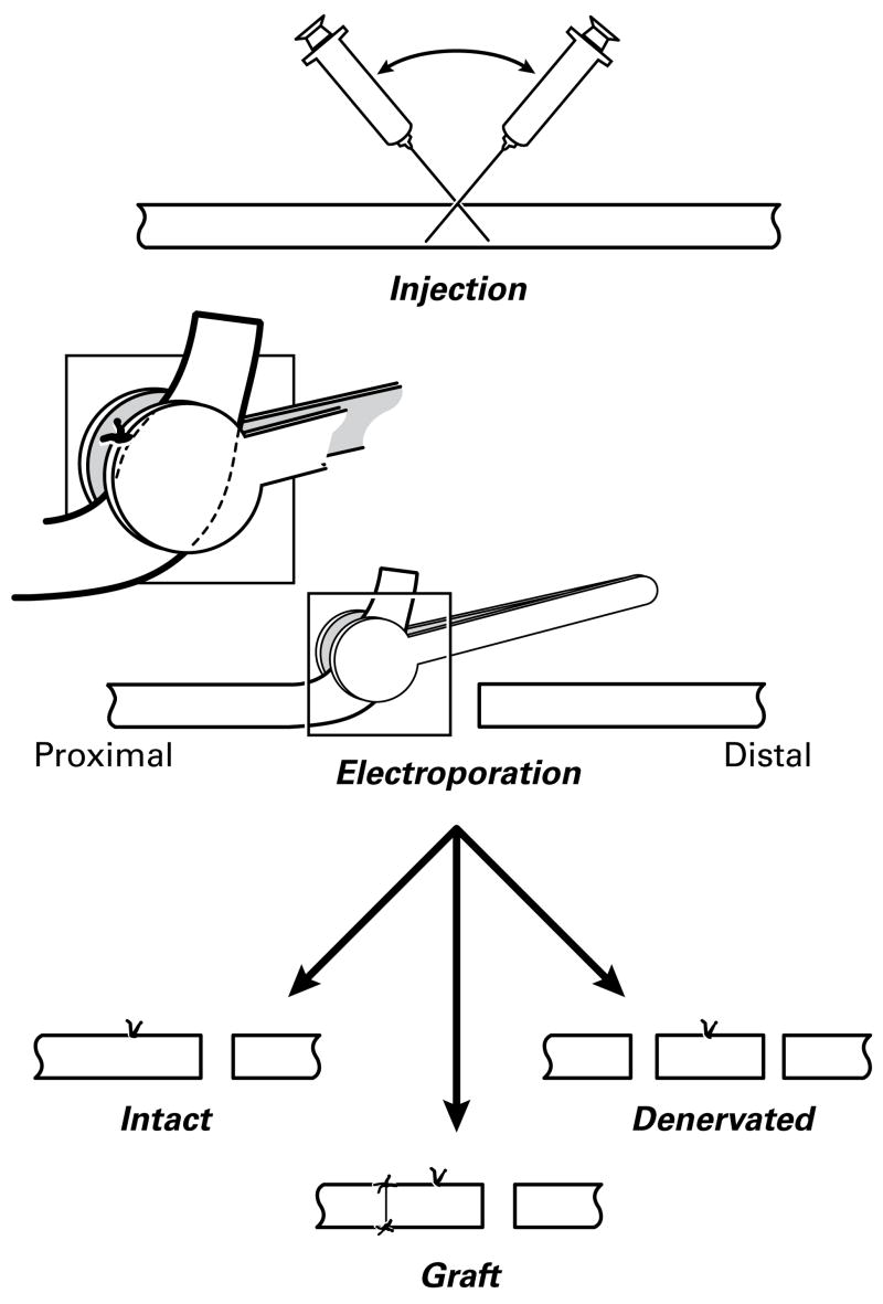
Figure 1.
Electroporation of rat tibial nerve. Proximal is to the left. After plasmid injection, the site of penetration is marked with a suture that is used as a landmark to center the electroporation paddles. The nerve is transected 8 mm distal to the suture and reflected to facilitate this process. It is then replaced in the wound without further manipulation (Intact group), or transected again 8mm proximal to the suture. This detached segment of nerve is then sutured back to the proximal stump so that it can be reinnervated (Graft group) or left detached (Denervated group). The Graft preparation was used both for the preliminary calibration studies as part of the long-term expression experiments.

