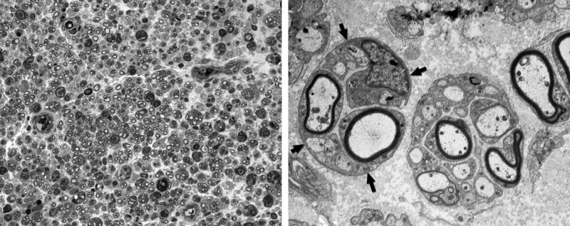
Figure 10.
Grafted nerve, 21 days after electroporation. Left: One micrometer plastic section, 100×. The nerve is populated with regenerating units, groups of regenerating axons contained within the basal lamina of Schwann cell tubes that have been cleared by Wallerian degeneration. Right: Electronmicrograph, 4,000×. Two regenerating units with fibers in various stages of myelination. The arrows outline the Schwann cell basal lamina that contains a single regenerating unit.

