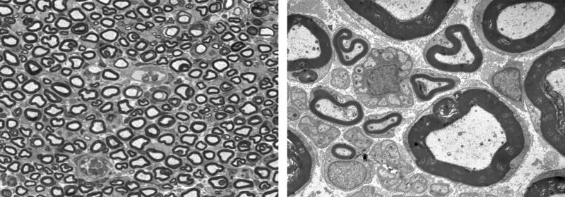
Figure 7.
Intact nerve, 3 days after electroporation. Left: One micrometer plastic section, 100×. No significant fiber loss is noted, and myelinated axons are closely packed. Right: Electronmicrograph, 4,000×. Myelinated and unmyelinated fibers are intact and there is sparse endoneurial space between axons.

