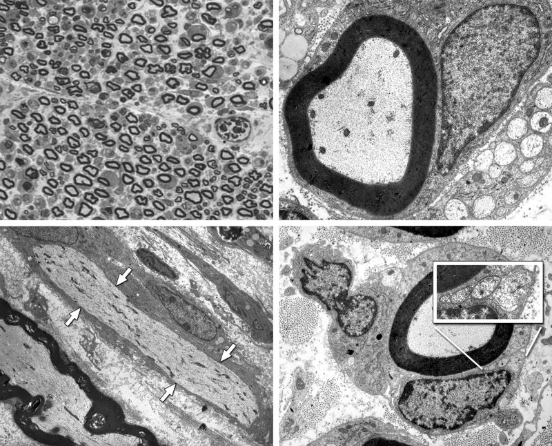
Figure 8.
Intact nerve, 7 days after electroporation. Upper Left: One micrometer plastic section, 100×. Endoneurial edema results in increased space between axons, and some myelinated profiles are lost. Upper Right: Electronmicrograph, 10,000×. Many myelinated axons and their accompanying Schwann cells are morphologically normal. Lower Left: Electronmicrograph of longitudinal section, 4,000×. The axon that runs from upper left to lower right, indicated by white arrows, is intact, but has undergone segmental demyelination. The axon at the lower left has maintained its myelin sheath. Lower Right: Many axons that remain myelinated are accompanied by unmyelinated sprouts within the same Schwann cell basal lamina (inset).

