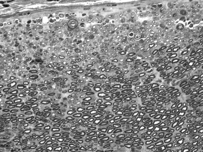
Figure 9.
Intact nerve, 14 days after electroporation. One micrometer plastic section, 100×, taken from the nerve that was damaged most severely by electroporation. Axonal loss and/or demyelination is prominent near the perineurium (upper edge), but more central portions of the nerve appear normal. Endoneurial edema has resolved as evidenced by the tight packing of myelinated axons.

