
Figure 3.
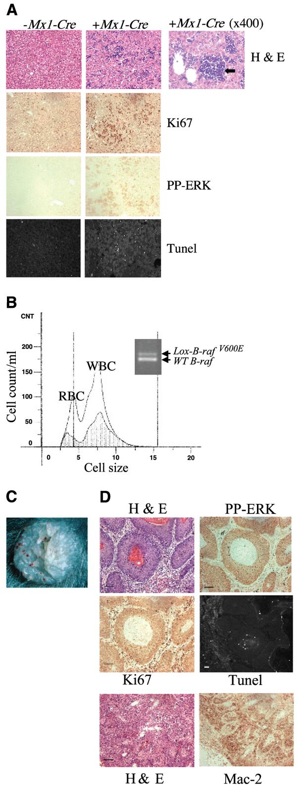
Analysis of tissues from mice resulting from LSL-B-rafV600E × Mx1-Cre intercrosses. A, histologic analysis of liver sections from LSL-B-rafV600E ± Mx1-Cre mice. Liver sections were either stained with H&E, with a Ki67 antibody, with a phospho-ERK antibody or processed for TUNEL analysis. Right, H&E section was photographed at higher magnification (×400) to indicate site of hemopoiesis (black arrow) in the mutant liver. Bar, 50 μm (the same for all sections) except for 25 μm (enlarged H&E section, right). B, analysis of bone marrow from LSL-B-rafV600E ± Mx1-Cre mice. Bone marrow cells were isolated from the femurs of LSL-B-rafV600E ± Mx1-Cre mice and analyzed by the Coulter counter. There are far fewer RBC and WBC in the bone marrow of LSL-B-rafV600E + Mx1-Cre mice (shaded area) than LSL-B-rafV600E − Mx1-Cre mice (clear area). PCR analysis of DNA from bone marrow of LSL-B-rafV600E + Mx1-Cre animal, indicating the presence of the Lox-B-rafV600E allele. C, photograph of histiocyte arising on skin of LSL-B-rafV600E + Mx1-Cre animal. D, histologic analysis of histiocyte sections from LSL-B-rafV600E ± Mx1-Cre mice. Sections were either stained with H&E, with antibodies for Ki67, phospho-ERK, or Mac-2, or processed for TUNEL analysis. Bar, 50 μm.