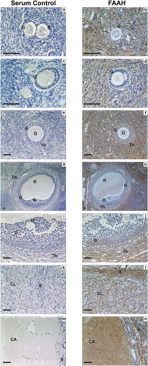
Figure 3. Immunohistochemical staining for FAAH.
Images on the left side of the panel represent the negative control (non-immune rabbit serum and images on the right are for FAAH. Images a and b are primordial follicle; c and d are primary follicles; e and f are secondary follicles; g and h are low power images of tertiary follicles; i and j are high power images of tertiary follicles, k and l are corpus luteum, and m and n are images of the corpus albicans. Granulosa cells (G), theca cell layers (Th), the oocyte (O) and follicular fluid (ff) demonstrated FAAH immunoreactivity as did lutein-granulosa cells of the corpus luteum (CL) and corpus albicans (CL), and septa (S). There was a gradation of staining in the granulosa cells of the tertiary follicle with greatest intensity in the mural cells (panel j). The images are representatives from at least two structures and taken at 50×, 200× or 400× magnification. Bar = 50 µm.

