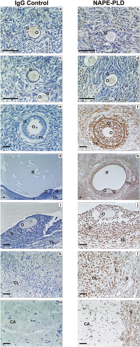
Figure 4. Immunohistochemical staining for NAPE-PLD.
Images in the left side of the panel represent the negative control (rabbit IgG) and images on the right are for NAPE-PLD. Images a, and b are primordial follicles, c and d are primary follicles, e and f are secondary follicles; g and h are low power images of tertiary follicles, i and j are high power images of tertiary follicles, k and i are images of corpus luteum, and m and n are images of corpus albicans. Granulosa cells (G), theca cell layers (Th), the oocyte (O) and follicular fluid (ff) demonstrated NAPE-PLD immunoreactivity as did lutein-granulosa cells of the corpus luteum (CL) and corpus albicans (CL) and septa (S). The images are representative from at least two structures and taken at 50×, 200×, or 400× magnification. Bar = 50 µm.

