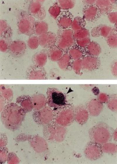
Figure 3.

In situ DNA PCR analysis of marrow or peripheral blood cells 24 hr posttransduction or from patients at various time intervals posttransplant. A Boehringer Mannheim kit was used for an in situ DNA PCR assay as described previously (16). The nuclei of the cells that are transduced turn blue, whereas those that are not transduced stay white. Controls that were run with every patient sample included cells known to be negative for the vector pVMDR-1 and the patient’s own pretransduction cells. The percent of cells positive in these negative controls (which is probably due to a primer independent repair incorporation reaction) was subtracted from the percent of cells positive for the blue intranuclear color in known negative cells (16). In situ DNA PCR analysis of negative control cells (A) and cells transduced by the stromal transduction method (B). The positive cells contain intensely staining blue nuclei.
