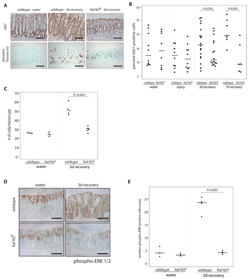
Figure 4. Raf is required for the epithelial hyperproliferative response following DSS-induced injury.
(A) Proliferating cells were detected by Ki67 and phospho-histone H3 staining in colon sections of wildtype and Raf KOIE mice during 3 day recovery from DSS-induced colitis. (B) The percentage of Ki67 positive cells in the midcolon are represented graphically. (C) The number of cells per hemicrypt are represented graphically. (D) Phospho-ERK staining of colon sections from control and Raf KOIE mice during 3 day recovery. Scale bars = 125μm. (E) The average number of phospho-ERK-positive cells per crypt are represented graphically. Horizontal lines represent the median.

