Fig. 8.
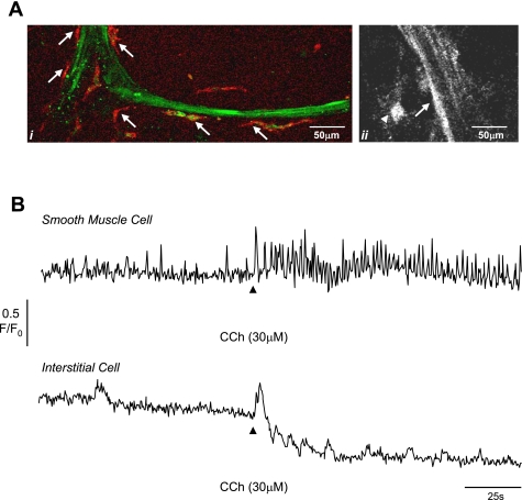
Ai: preparation showing smooth muscle bundle loaded with fluo 4AM (green) and labeled with anti-c-Kit (red) showing the interstitial cells (arrows) on the edge of the bundle. Aii: smooth muscle bundle and interstitial cell to left border of bundle. B: intensity time series from smooth muscle cell (arrow in Aii) and interstitial cell (arrow head in Aii) showing the effect of 30 μM carbachol. Carbachol increased the frequency of spontaneous transients in the smooth muscle cell and caused an additional increase in intracellular Ca2+ in the interstitial cell followed by several oscillations. The reduction in baseline in the interstitial cell record is due to movement of the tissue. This record is typical of 5 experiments described in the text.

