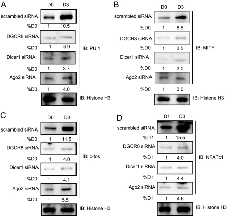
FIGURE 2.
Expression of transcription factors in osteoclast differentiation. Infected cells were treated with RANKL (50 ng/ml) for 3 days, and nuclear extracts were analyzed by immunoblotting with antibodies against PU.1 (A), MITF (B), c-Fos (C), and NFATc1 (D). Quantitative image analysis of the protein expression levels (values below bands) was normalized to histone H3. IB, immunoblotting.

