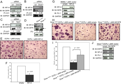
FIGURE 7.
A negative effect of NFI-A on M-CSFR expression. A-D, G, and J, NFI-A, M-CSFR, and PU.1 levels in infected-osteoclast precursors and Dicer-null osteoclast precursors were analyzed by immunoblotting using whole cell extracts. Quantitative image analysis of these protein expression levels were normalized to GAPDH. scr, scrambled. IB, immunoblotting. E, F, H, and I, infected osteoclast precursors were replated in 24-well plates, and the cells were treated with RANKL (50 ng/ml) and M-CSF-conditioned medium (1:20). After 5 days, the cells were then fixed and stained for TRAP, and the number of TRAP-positive multinucleated cells was scored. Similar findings were obtained in four independent sets of experiments. OCP, osteoclast precursors.

