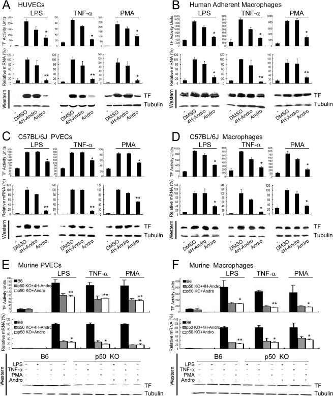
FIGURE 1.
Andro treatment and p50 knock-out inhibit TF activity and expression in stimulated human and murine endothelial cells and monocytes/macrophages. A–D, HUVECs (A), human adherent macrophages (B), C57BL/6J PVECs (C), and C57BL/6J macrophages (D) were stimulated with LPS, TNF-α, and PMA, in the presence of buffer (-), DMSO (a solvent control), 4H-Andro (an inactive structural analog), and Andro. Cell lysates were measured for TF activity (upper panels) and TF expression (lower panels). Results are presented as either the mean ± S.D. value of triplicate measurements of three independent experiments (upper panels,*, p < 0.05; **, p < 0.01 versus DMSO group) or a representative of three independent experiments (lower panels). Although 4H-Andro appeared to partially inhibit TF activity and mRNA induced by TNF-α and PMA, these differences were not statistically significant (p > 0.05). E and F, TF activity and expression in stimulated endothelial cells and macrophages isolated from p50 null mice. PVECs (E) and macrophages (F) were isolated from B6 mice and p50 null mice. Following treatment with LPS, TNF-α, and PMA in the absence or presence of 4H-Andro and Andro, the activity (upper panels) and expression (lower panels) of TF were measured. Results are presented as either the mean ± S.D. value of triplicate measurements of three independent experiments (upper panels,*, p < 0.05; **, p < 0.01 versus B6 group) or a representative of three independent experiments (lower panels).

