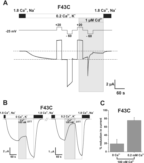
FIGURE 4.
Cd2+ block of F43C hemichannels is promoted during closure by hyperpolarization and extracellular Ca2+. A, an oocyte expressing F43C was voltage clamped at -25 mV and extracellular Ca2+ was lowered to 0.2 mm in a KCl solution. A pair of 30-s voltage steps were applied, one depolarizing to +20 mV to place F43C channels in the open state and one hyperpolarizing to -60 mV to promote closure of the loop gate. Application of 1 μm Cd2+ at +20 mV (shaded area) produced little or no block of the outward current. However, subsequent hyperpolarization to -60 mV in the continued presence of Cd2+ caused a rapid reduction in the current, indicating that the introduced cysteines move during loop gating and form metal bridges. B, shown are membrane currents in an oocyte voltage clamped at -30 mV. Perfusing a KCl solution containing no added Ca2+ (0 Ca2+, K+) produced a substantial inward current. Application of 100 nm Cd2+ produced a modest reduction in current of ∼20%. Brief application of DTT increased current beyond that in 0 mm Ca2+ alone indicating there was some modest lock-up. Changing a KCl solution containing 0.2 mm Ca2+ (0.2 Ca2+, K+) decreased the current (note change in scale) and addition of the same 100 nm concentration of Cd2+ now produced a substantial reduction in current of ∼75%. Brief application of DTT similarly increased current beyond levels in 0.2 mm Ca2+ alone. C, bar graph summarizing the effect of 100 nm Cd2+ applied at -30 mV in 0 mm Ca2+ (n = 5) and 0.2 mm Ca2+ (n = 8). Bars represent the mean ± S.E. of block.

