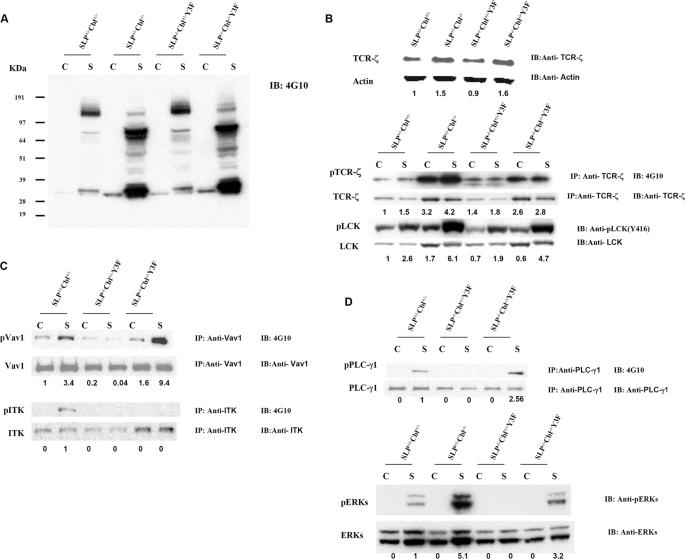
FIGURE 4.
Cbl inactivation reverses selective signaling defects in SLP76-/-Y3F thymocytes. DP thymocytes were sorted from SLP76+/-Cbl+/- (wild type), SLP76+/-Cbl-/- (Cbl-/-), SLP76-/-Cbl+/-Y3F (SLP76-/-Y3F), and SLP76-/-Cbl-/-Y3F mice by flow cytometry and were unstimulated or stimulated with anti-CD3 Abs for 5 min at 37 °C. Protein lysates were prepared from DP thymocytes. A, protein lysates were immunoblotted (IB) with anti-phosphotyrosine Ab (4G10). B, protein lysates were immunoblotted (IB) with anti-TCR-ζ, anti-actin, anti-Lck, or anti-phospho(P)-lck antibody or were immunoprecipitated (IP) with anti-TCR-ζ and immunoblotted with anti-phosphotyrosine Ab (4G10) and anti-TCR-ζ,(upper panels). C, protein lysates were immunoprecipitated with anti-Vav1 and immunoblotted with anti-phosphotyrosine Ab (4G10) and anti-Vav1 (upper panels) or were immunoprecipitated with anti-ITK and immunoblotted with 4G10 and anti-ITK (lower panels). D, protein lysates were immunoprecipitated with anti-PLCγ1 and immunoblotted with anti-phosphotyrosine (4G10) or anti-PLCγ1(upper panels), or lysates were immunoblotted with anti-phospho-ERKs or anti-ERK (lower panels). Lane C indicates control cells without stimulation; lane S indicates cells were stimulated with 10 μg/ml of anti-mouse CD3 antibodies for 5 min.

