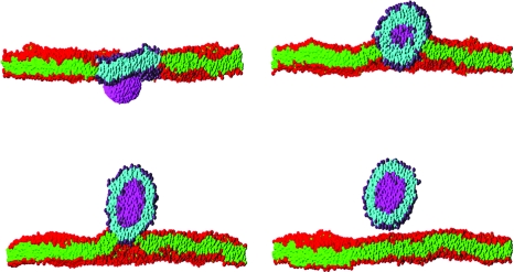
Figure 1. Sequence of snapshots of a cross section through a rigid nanoparticle (magenta) translocating across a planar lipid bilayer membrane immersed in solvent (invisible for clarity).
The times of the snapshots are 130 ns, 650 ns, 1.3 μs and 2.8 μs labeled top to bottom by row. The membrane contains 7973 lipids and spans the 50×50 nm2 simulation box. It initially contains a circular patch of 480 molecules of one lipid type (purple headgroups and cyan tails) surrounded by 7493 of the second lipid type (red headgroups and green tails). Both types have identical molecular architecture, but their interactions are chosen so that they phase separate in the membrane. The nanoparticle has approximate dimensions 8×6×6 nm3, and its interactions are chosen so that it prefers to adhere to the patch lipid headgroups rather than the other lipid type. The combination of the adhesion of the nanoparticle to the patch, which induces it to curve, and the line tension of the patch boundary drives the translocation process. The snapshots are taken from a dissipative particle dynamics simulation of the author, and are rendered using the PovRay software (www.povray.org).

