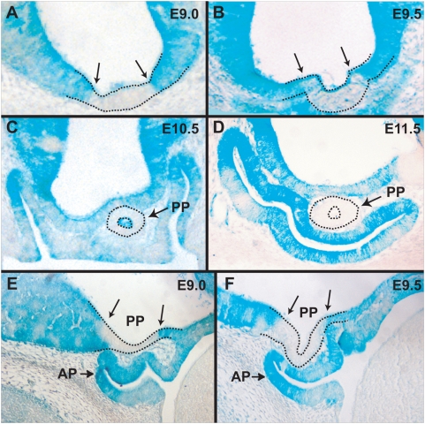
Figure 3. Formation of the posterior pituitary in chimeric embryos.
(A) Section through an E9.0 chimeric embryo demonstrating a region free of Rx-deficient cells in the ventral hypothalamus. Two arrows indicate the sharp boundaries between the wild type cells (white) and Rx−/− cells (blue). (B) Section through an E9.5 chimeric embryo demonstrating that the evaginating posterior pituitary does not contain any Rx-deficient cells. Two arrows indicate the sharp boundaries between the wild type cells and Rx−/− cells. (C) Section through an E10.5 chimeric embryo showing that the tubular posterior pituitary is devoid of Rx−/− cells. (D) Section through an E11.5 chimeric embryo demonstrating that the posterior pituitary is devoid of Rx−/− cells. (E) Sagittal section of an E9.0 chimeric embryo visualizing the Rx−/− cell free region in the ventral hypothalamus. (F) Sagittal section of an E9.5 chimeric embryo showing that the evaginating posterior pituitary does not contain Rx−/− cells. AP - anterior pituitary, PP – posterior pituitary.

